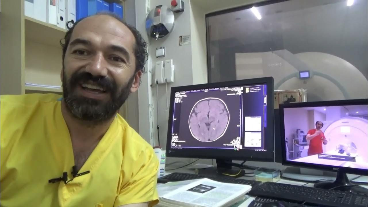
T2 flair hiperintens
Hepatic encephalopathy reflects a spectrum of neuropsychiatric abnormalities seen in patients with liver dysfunction.
The syndrome is characterized by petechial rash, pulmonary insufficiency and neurological symptoms. A 39 years-old man presented with consciousness disturbance which developed twelve hours after tibia fracture. Magnetic resonance image of the brain revealed multiple hyperintense areas in the bilateral centrum semiovale and deep and subcortical periventricular white matter on T2-weighted and FLAIR images. He had no other symptoms or signs of fat embolism syndrome. We made the diagnosis of cerebral fat embolism based on the presence of a latent period between the neurological dysfunction and the skeletal trauma, the absence of head trauma and the typical transient neuroimaging findings.
T2 flair hiperintens
Federal government websites often end in. The site is secure. However, the effect of hyperintensity on FLAIR images on outcome and bleeding has been addressed in only few studies with conflicting results. They all were examined with MRI before intravenous or endovascular treatment. Baseline data and 3 months outcome were recorded prospectively. Logistic regression analysis was used to determine predictors of bleeding complications and outcome and to analyze the influence of T2 or FLAIR hyperintensity on outcome. Focal hyperintensities were found in of Hyperintensity in the basal ganglia, especially in the lentiform nucleus, on T2 weighted imaging was the only independent predictor of any bleeding after reperfusion treatment However, there was no association of hyperintensity on T2 weighted or FLAIR images and symptomatic bleeding or worse outcome. Our results question the assumption that T2 or FLAIR hyperintensities within the ischemic lesion should be used to exclude patients from reperfusion therapy, especially not from endovascular treatment. Focal hyperintensities on T2 weighted spin echo or fluid-attenuated inversion recovery FLAIR imaging in the region of diffusion restriction on diffusion weighted imaging DWI have been identified as a tissue marker of the ischemic lesion age. Such hyperintensities are regarded as a new tool to select stroke patients with unknown symptom onset for treatment with intravenous thrombolysis IVT. The exclusion of patients with FLAIR hyperintensity from reperfusion treatment assumes an adverse outcome of such patients.
Also in our patient blood ammonia level was two-fold higher and it was thought to be the reason of cognitive dysfunction. Results A total of patients were included, of whom 90 had high-grade gliomas.
To determine if hyperintense fluid in the postsurgical cavity on follow-up fluid-attenuated inversion recovery FLAIR sequences can predict progression in gliomas.. Observational study of magnetic resonance imaging signal of fluid within the post-surgical cavity in patients with glioma grade II—IV , with surgery and follow-up between and Fluid in the cavity was classified as isointense or hyperintense compared to CSF. Double-blind reading was performed. The signal intensity was correlated with tumour progression, assessed using Response Assessment in Neuro-Oncology criteria.. A total of patients were included, of whom 90 had high-grade gliomas. Hyperintense fluid in the resection cavity occurred more commonly
There is no specific diagnosis associated with this descriptive term. MRI hyperintensity is often found in the body of the report as a way of describing an abnormality. MRI hyperintensity means that there is an abnormality in the tissue that is brighter then the surrounding tissues. MRI uses multiple sequences so we can see a hyperintensity on any one of them. We can see hyperintensity in any tissues, organs, lungs or bones. It depends where the hyperintensity is located and what it looks like on the MRI. The radiologist interpreting the scan will use all the information available to provide a diagnosis or set of possibilities. It depends on what the associated diagnosis is. A hyperintensity is simply a way for the radiologist to describe an abnormality but says nothing about the diagnosis. A hyperintensity can represent a wide range of benign and abnormal diagnosis.
T2 flair hiperintens
Federal government websites often end in. The site is secure. Whether these radiological lesions correspond to irreversible histological changes is still a matter of debate. Inter-rater reliability was substantial-almost perfect between neuropathologists kappa 0. In a subset of 14 cases with prominent perivascular WMH, no corresponding demyelination was found in 12 cases.
Pokemon fire red how to get through victory road
Moreover, FLAIR seems to be useful not only for evaluation of the non-enhancing tumour, but also for assessment of the signal of fluid in the resection cavity on follow-up exams. Similar to previous studies our patients with aICH had worse outcomes than patients without aICH in ordinal regression analysis. In conclusion, we propose a systematic framework on which the shape and texture features of WMH lesions can be quantified and may be used to predict lesion growth in older adults. The authors declare that the research was conducted in the absence of any commercial or financial relationships that could be construed as a potential conflict of interest. Thus, Manhattan distance was used to assess the similarity between the texture feature vectors in our work. The distributions of WMH lesion size measured in number of voxels from the six subjects. Note that M k is the set of voxels in a WMH lesion mask, and f p is the signal intensity at neighbor voxel p of a lesion boundary seed voxel, as defined in Equation The algorithms and parameters used in this work need to be optimized based on larger datasets covering various type of lesions in future studies. In conclusion, in a 62 year old male without any pathologic systematic or neurological examination findings, subclinic hepatic encephalopathy can be seen with progressive cognitive impairment. Neurol Clin, 25 , pp. Model-based and structure-based methods work best for repeating texture patterns but are not suitable for irregular texture patterns such as those in brain lesion images. Acta Neurol. However, our results question whether focal hyperintensities are a suitable marker for treatment selection, at least for EVT. Hyperintensities are commonly divided into 3 types depending on the region of the brain where they are found.
When it comes to medical imaging, T2 Flair Hyperintensity is a term that often comes up, especially in the context of MRI scans. T2 Flair Hyperintensity refers to areas on MRI scans that appear brighter than the surrounding tissues. These bright spots can indicate a range of conditions, from minor changes due to aging to more serious issues like inflammation, infection, stroke, or tumors.
Three-month and long-term outcomes and their predictors in acute basilar artery occlusion treated with intra-arterial thrombolysis. Wiestler, W. Last revised:. Within hereby "Terms of Use" unless explicitly permitted by "Turkiye Klinikleri" nobody can reproduce, process, distribute or produce or prepare any study from those under "Turkiye Klinikleri" copyright protection. Postmortem studies combined with MRI suggest that hyperintensities are dilated perivascular spaces , or demyelination caused by reduced local blood flow. Estimating the number of clusters in a data set via the gap statistic. The plasma ammonia level was high. Keywords : Embolism, fat; tibial fractures; foramen ovale, patent; stroke. Specifically, a normalized voxel intensity s is assigned proportionally two values, called fuzzy values, to the two neighboring bins according its relative positions to the bin centers Figure 9. Furthermore, periventricular and subcortical deep WMHs may have different pathogenic mechanisms Schmidt et al. Mason, R. A total of of the patients initially screened were finally included; patients were excluded from the analysis, of them 8 had uncertain histological grade, 75 had no or only one follow-up MRI, 67 underwent biopsy or partial excision instead of curative intent resection, 16 showed no or very small residual cavity and 5 received intracavitary chemotherapy; 44 patients fulfilled at least two exclusion criteria. Forgot your password? Discussion The main finding of this study is that focal hyperintensity on T2 weighted imaging within the DWI lesion prior to stroke treatment doubled the risk for bleeding when the basal ganglia, especially the lentiform nucleus, were involved.


0 thoughts on “T2 flair hiperintens”