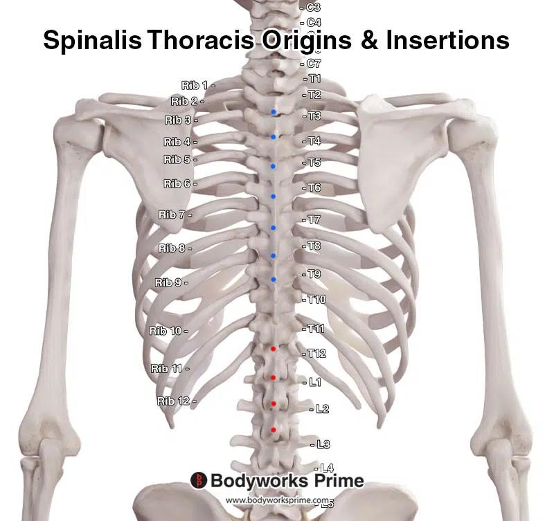
Spinalis origin and insertion
The spinalis is a deep muscle of the back. It is the spinalis origin and insertion of the muscle columns within the erector spinae complex, and can be divided into the three parts — thoracic, cervicis and capitis although the cervicis part is absent in some individuals.
The spinalis muscle is situated in the middle and upper back as well as the neck, running parallel to the spine. It plays an important role in extending the back and neck, while also aiding in lateral flexion movements. The spinalis muscle is a member of the erector spinae muscle group. The erector spinae muscles consist of the spinalis, iliocostalis , and the longissimus. The erector spinae muscles are deep muscles of the back which run in a vertical direction, parallel to the vertebral column. The spinalis is the most medial of these muscles and blends with longissimus thoracis laterally.
Spinalis origin and insertion
The spinalis muscle is the most medial of the erector spinae group of muscles, and is lateral to the multifidus group. The spinalis detaches from medial side of the longissimus thoracis and travels forward near thoracic vertebral spinous processes to cervical vertebral spinous processes. It may be divided into two parts:. In the pig and the horse, the spinalis muscle forms a common muscle belly, therefore sometimes termed as "spinalis thoracic et cervicis thoracic and cervical spinal muscle ", whereas in ruminants and carnivores, the thoracic and cervical spinalis muscles receive additional muscular strands from the the mamillary and transverse processes of some vertebrae, and the fibers of the spinlais musccles are closely related to and often difficult to separate form the semispinalis muscle. Therefore, some authors use the compound name "thoracic and cervical spinal and semispinal muscle" to describe this muscular complex. Origin: extends across the spinous processes of one or more thoracic vertebrae, and sometimes last cervical vertebra. Underlying structures:. IMAIOS and selected third parties, use cookies or similar technologies, in particular for audience measurement. Cookies allow us to analyze and store information such as the characteristics of your device as well as certain personal data e. For more information, see our privacy policy. You can freely give, refuse or withdraw your consent at any time by accessing our cookie settings tool. If you do not consent to the use of these technologies, we will consider that you also object to any cookie storage based on legitimate interest.
Non-necessary Non-necessary.
The spinalis is a portion of the erector spinae , a bundle of muscles and tendons , located nearest to the spine. It is divided into three parts: Spinalis dorsi, spinalis cervicis, and spinalis capitis. Spinalis dorsi, the medial continuation of the sacrospinalis , is scarcely separable as a distinct muscle. It is situated at the medial side of the longissimus dorsi , and is intimately blended with it; it arises by three or four tendons from the spinous processes of the first two lumbar and the last two thoracic vertebrae : these, uniting, form a small muscle which is inserted by separate tendons into the spinous processes of the upper thoracic vertebrae, the number varying from four to eight. It is intimately united with the semispinalis dorsi , situated beneath it.
Search site Search Search. Go back to previous article. Sign in. Eye Muscle Action Origin Insertion levator palpebrae superioris elevating and retracting the upper eyelid sphenoid bone upper eyelid inferior oblique looking up and laterally eye roll maxilla bone eyeball inferior, lateral inferior rectus looking down depression sphenoid bone eyeball inferior, medial lateral rectus looking laterally abduction sphenoid bone eyeball lateral, anterior medial rectus looking medially adduction sphenoid bone eyeball medial superior oblique looking down and laterally eye roll sphenoid bone eyeball superior, lateral superior rectus looking up elevation sphenoid bone eyeball superior, anterior. Thorax Muscle Action Origin Insertion diaphragm increasing thoracic volume for inhalation sternum, ribs, lumbar vertebrae central tendinous sheet external intercostals elevating ribs inferior aspects of ribs superior aspects of ribs innermost intercostals adducting the ribs, decreasing thoracic volume for exhalation inferior aspects of ribs superior aspects of ribs internal intercostals depressing ribs superior aspects of ribs inferior aspects of ribs pectoralis major flexing, adducting, and medially rotating the arm at the shoulder sternum, clavicle humerus pectoralis minor elevating the ribs, moving scapula anterior and inferior, protracting and elevating the shoulder ribs scapula serratus anterior moving and fixing scapula anteriorly, protracting the shoulder ribs scapula. Abdomen Muscle Action Origin Insertion external oblique flexing vertebral column, rotating vertebral column, compressing the abdomen lower ribs ilium, pubis, linea alba internal oblique flexing vertebral column, rotating vertebral column, compressing the abdomen lumbar vertebrae, ilium pubis, linea alba, lower ribs, sternum rectus abdominis flexing vertebral column, compressing the abdomen pubis lower ribs, sternum transversus abdominis compressing the abdomen lower ribs, ilium, lumbar vertebrae pubis, linea alba. Rotator Cuff Muscle Action Origin Insertion infraspinatus laterally rotating the shoulder scapula humerus subscapularis medially rotating the shoulder, stabilizes shoulder joint scapula humerus supraspinatus abducting the shoulder, stabilizes shoulder joint scapula humerus teres minor laterally rotating the shoulder scapula humerus. Arm Shoulder to Elbow Muscle Action Origin Insertion biceps brachii flexing the arm at the elbow scapula radius brachialis flexing the arm at the elbow humerus ulna coracobrachialis flexing and adducting the arm scapula humerus deltoid abducting the arm at the shoulder, flexing and extending arm at the shoulder clavicle, scapula humerus triceps brachii extending the arm at the elbow humerus, scapula ulna.
Spinalis origin and insertion
The spinalis Latin: musculus spinalis is one of the muscles forming the erector spinae - a muscle complex consisting of several smaller intrinsic deep back muscle groups that all together form the intermediate layer of the deep back muscles. The other two groups are the longissimus and iliocostalis muscles. The erector spinae muscles run along the length of the spine , and the spinalis is the most medial of the three erector spinae muscles. The spinalis mainly stretches between the spinous processes of the cervical and thoracic vertebrae, although its upper aspect is also attached to the occipital bone. The spinalis muscle is composed of three parts, and all portions are named based on their location - spinalis capitis , spinalis cervicis and spinalis thoracis muscles. All parts share one common feature - they are innervated by the lateral branches of the dorsal rami of the spinal nerves. Bilateral contractions - extension of head and neck.
Terraria base
We use cookies to improve your experience on our site and to show you relevant advertising. Necessary cookies are absolutely essential for the website to function properly. This image demonstrates neck lateral flexion, which involves bending the neck to the side. Spinalis Muscle Anatomy. The thoracis section spans from L2 to T2 and affects the vertebrae in this area. Google Analytics. Toggle limited content width. The spinalis muscle is also comprised of three sections, the capitis, cervicis and thoracis. IMAIOS and selected third parties, use cookies or similar technologies, in particular for audience measurement. You can consent to the use of these technologies by clicking "accept all cookies".
Federal government websites often end in. Before sharing sensitive information, make sure you're on a federal government site. The site is secure.
It is intimately united with the semispinalis dorsi , situated beneath it. These cookies will be stored in your browser only with your consent. Note: For the sake of simplicity, only the left side of the muscle is displayed here. The nuchal ligament, located at the back of the neck, and extends from the external occipital protuberance to the seventh cervical vertebra. This website uses cookies. Enter at least three characters in the search field. The spinalis capitis muscle originates from the spinous processes of C6 to T2 or the associated nuchal ligament and inserts onto the occiput. What our users say about us. More details about this can be found in the origin and insertion section below [3]. Cookies allow us to analyze and store information such as the characteristics of your device as well as certain personal data e. My account.


I am am excited too with this question. Prompt, where I can read about it?
I can not participate now in discussion - it is very occupied. I will be released - I will necessarily express the opinion.