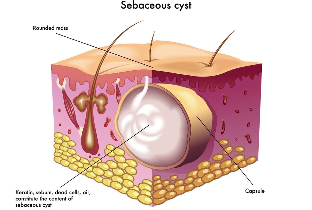
Pilar cyst diagram
Federal government websites often end in.
DermNet provides Google Translate, a free machine translation service. Note that this may not provide an exact translation in all languages. Home arrow-right-small-blue Topics A—Z arrow-right-small-blue Trichilemmal cyst. Advisor to the content: Dr. Copy Editor: Clare Morrison.
Pilar cyst diagram
Pilar cysts grow around hair follicles and usually appear on the scalp. They are yellow or white and form small, round, or dome-shaped bumps. They grow slowly and may disappear on their own. In some cases, a doctor may remove them. A cyst is a small lump filled with fluid. They form under the skin. Cysts are very common and usually have no symptoms or side effects. There are three main types of cysts: epidermoid , meibomian chalazion , and pilar cysts. Many pilar cysts heal without treatment if a person is careful not to damage the skin. A surgeon is usually able to remove a cyst easily. However, sometimes even after removal, the cyst may reappear. A cyst will appear as a small, round, or dome-shaped bump.
Hidden categories: Articles with short description Short description is different from Wikidata Use dmy dates from October Frequently asked questions.
A trichilemmal cyst or pilar cyst is a common cyst that forms from a hair follicle , most often on the scalp , and is smooth, mobile, and filled with keratin , a protein component found in hair , nails , skin , and horns. Trichilemmal cysts are clinically and histologically distinct from trichilemmal horns, hard tissue that is much rarer and not limited to the scalp. Trichilemmal cysts may be classified as sebaceous cysts , [6] although technically speaking are not sebaceous. Medical professionals have suggested that the term "sebaceous cyst" be avoided since it can be misleading. Trichilemmal cysts are derived from the outer root sheath of the hair follicle. Their origin is currently unknown, but they may be produced by budding from the external root sheath as a genetically determined structural aberration.
Pilar Cysts, also known as Trichilemmal Cysts, are keratin-filled bumps that grow on the skin. How is this bump different from the many others that are found on the body? This cyst is filled with keratin, the specialized protein that is found in hair and nails. We shed our skin everyday. When the skin sheds into the pocket, the keratin accumulates and the cyst develops and grows. Adam Mamelak , board-certified dermatologist and skin surgeon in Austin.
Pilar cyst diagram
Pilar cysts tend to form on the scalp where keratin builds up in the lining of your hair follicles. They are typically harmless. Pilar cysts are flesh-colored bumps that can develop on the surface of the skin. You may be able to identify some of the characteristics of pilar cysts on your own, but you should still see your doctor for an official diagnosis. Keep reading to learn more about how these cysts present, whether they should be removed, and more. Pilar cysts grow within the surface of your skin. Although 90 percent of pilar cysts occur on the scalp, they can develop anywhere on the body. Other possible sites include the face and neck.
David garrett izmir
Clin Exp Reprod Med. Search term. Classification D. Cysts often heal on their own. In rare cases, this leads to formation of a tumor, known as a proliferating trichilemmal cyst. April Perioral dermatitis Granulomatous perioral dermatitis Phymatous rosacea Rhinophyma Blepharophyma Gnathophyma Metophyma Otophyma Papulopustular rosacea Lupoid rosacea Erythrotelangiectatic rosacea Glandular rosacea Gram-negative rosacea Steroid rosacea Ocular rosacea Persistent edema of rosacea Rosacea conglobata variants Periorificial dermatitis Pyoderma faciale. Medical condition. Pilar cysts are lined by thick capsules containing small layers of cuboidal, dark-staining basal epithelial cells in a palisade arrangement without an obvious intercellular gap. Retrieved 4 March Home arrow-right-small-blue Topics A—Z arrow-right-small-blue Trichilemmal cyst. Full name.
A pilar cyst is a common benign cyst usually found on the scalp.
Basaloid follicular hamartoma Folliculosebaceous cystic hamartoma Folliculosebaceous-apocrine hamartoma. Sequelae of trichilemmal cysts include inflammation, infection and malignant transformation which is rare. Recurrences and metastases have been observed. The pilar cyst rate of growth is very slow; it takes several years to grow to a big size. Reassurance of the patient is very important. Pilar cysts also tend to be more common in women than in men. It is not always necessary to remove a cyst. Others prefer a more conservative approach. Pilar cysts occur most commonly in middle-aged women. Pilar cyst Image from Dr Calum Lyon. Philadelphia: Saunders. Related Coverage. Wikimedia Commons. Treatments Your medical professional may: Cut into incise and drain the keratin and other material inside the cyst. N Am J Med Sci.


Excellent idea and it is duly
It is remarkable, it is rather valuable answer
It seems brilliant phrase to me is