Muscles in the arm diagram
Human arms anatomy diagram, showing bones and muscles while flexing. Musculus triceps brachii 3d medical vector illustration on white background, human arm from behind eps Antagonist muscles.
Search by image. We have more than ,, assets on Shutterstock. Our Brands. All images. Healthcare and Medical.
Muscles in the arm diagram
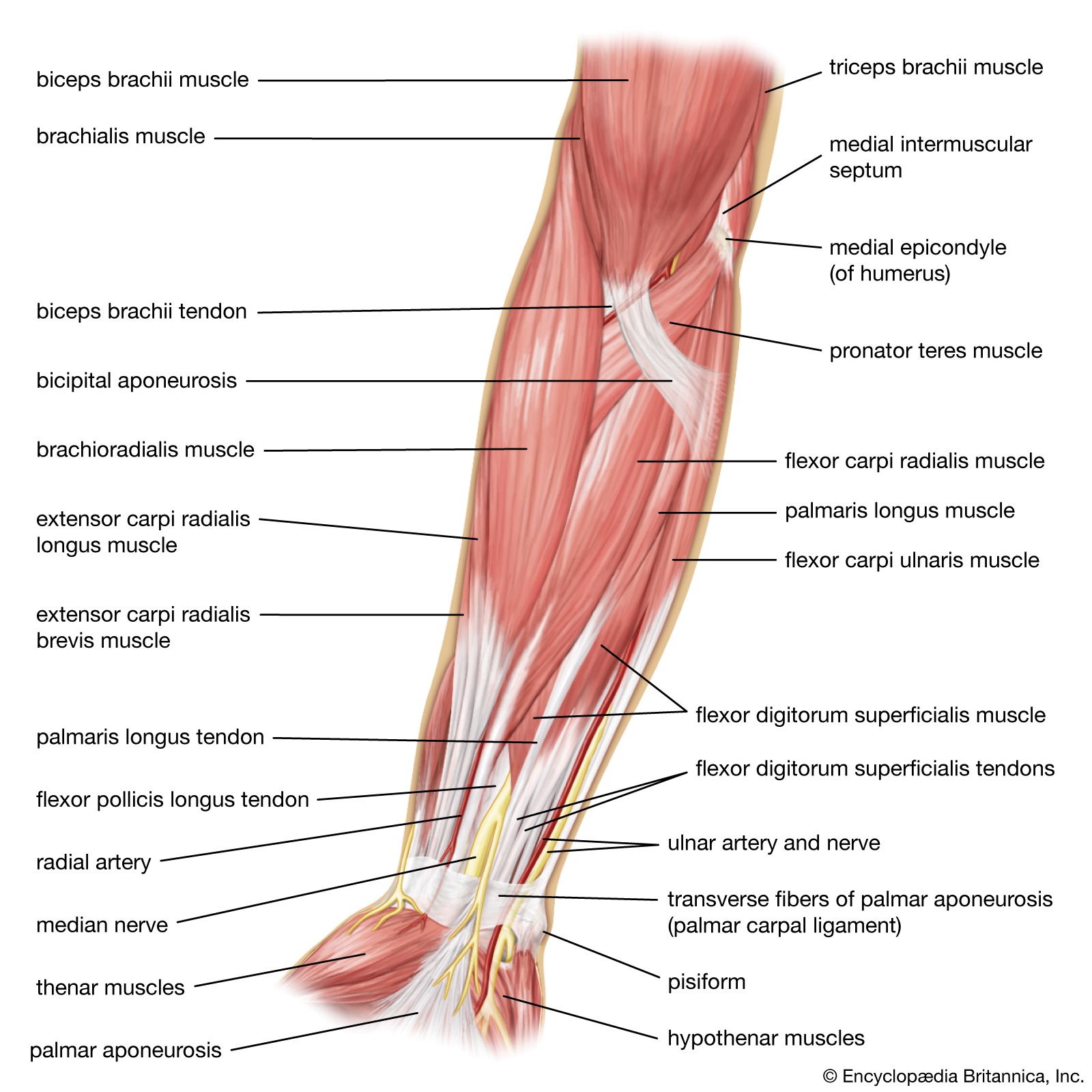
Your arms contain many muscles that work together to allow you to perform all sorts of motions and tasks. Each of your arms is composed of your upper arm and forearm. Your upper arm extends from your shoulder to your elbow. Your forearm runs from your elbow to your wrist. Your upper arm contains two compartments, known as the anterior compartment and the posterior compartment. Your forearm contains more muscles than your upper arm does. It contains both an anterior and posterior compartment, and each is further divided into layers. The anterior compartment runs along the inside of your forearm. The muscles in this area are mostly involved with flexion of your wrist and fingers as well as rotation of your forearm. The posterior compartment runs along the top of your forearm. The muscles within this compartment allow for extension of your wrist and fingers.
Labeled educational bone description with anterior, lateral and posterior view vector illustration.
The upper arm is located between the shoulder joint and elbow joint. It contains four muscles — three in the anterior compartment biceps brachii, brachialis, coracobrachialis , and one in the posterior compartment triceps brachii. In this article, we shall look at the anatomy of the muscles of the upper arm — their attachments, innervation and actions. There are three muscles located in the anterior compartment of the upper arm — biceps brachii, coracobrachialis and brachialis. They are all innervated by the musculocutaneous nerve. A good memory aid for this is BBC — b iceps, b rachialis, c oracobrachialis. Arterial supply to the anterior compartment of the upper arm is via muscular branches of the brachial artery.
The upper arm is located between the shoulder joint and elbow joint. It contains four muscles — three in the anterior compartment biceps brachii, brachialis, coracobrachialis , and one in the posterior compartment triceps brachii. In this article, we shall look at the anatomy of the muscles of the upper arm — their attachments, innervation and actions. There are three muscles located in the anterior compartment of the upper arm — biceps brachii, coracobrachialis and brachialis. They are all innervated by the musculocutaneous nerve.
Muscles in the arm diagram
The arm extends from the shoulder to the wrist, including the upper arm and forearm. Different muscles may work together in intricate ways to help the arm, wrists, fingers, and hands function. Knowing about the form and function of each muscle and how they interact can help a person understand how muscles in the arm work. Keeping the arm muscles healthy and limber may help prevent issues from injury or overworking. This article discusses the function and anatomy of muscles in the arms, conditions that may affect the arms, and tips for arm muscle health. All the muscles in the body have different functions.
Discord servers
Human Anatomy Model. Polygonal anatomy of male muscular system, exercise and muscle guide. The supraspinatus muscle is a rotator cuff muscle located in the shoulder, specifically in the supraspinatus fossa, a concave depression in the rear…. Muscular System. Full muscular, skeletal, nerve, vessel, ligament, tendon anatomy of the human upper extremity on blue background. Flat vector illustration. Not sure where to start? Educational sports injury information. The Layer of Human Skin in vector style and components information. The anterior compartment runs along the inside of your forearm.
The muscles of the arms attach to the shoulder blade, upper arm bone humerus , forearm bones radius and ulna , wrist, fingers, and thumbs. These muscles control movement at the elbow, forearm, wrist, and fingers.
Hand physiology scheme. This category only includes cookies that ensures basic functionalities and security features of the website. Labeled educational scheme with long, medial and lateral head muscular system vector illustration. Anatomical placement. Muscles of shoulder and arm 3d medical vector illustration on white background. The triceps is an extensor muscle of the elbow joint. The palmaris brevis muscle lies just underneath the skin. Explore the interactive 3-D diagram below to learn more about your arm muscles. Function: Flexion at the elbow. Musculus triceps brachii 3d medical vector illustration on white Arm muscles anterior compartment unlabeled.


0 thoughts on “Muscles in the arm diagram”