Mesenchyme
Thank mesenchyme for visiting nature. You are using a browser version with limited support for CSS. To obtain the best experience, mesenchyme, we recommend you use a more up to mesenchyme browser or turn off compatibility mode in Internet Explorer.
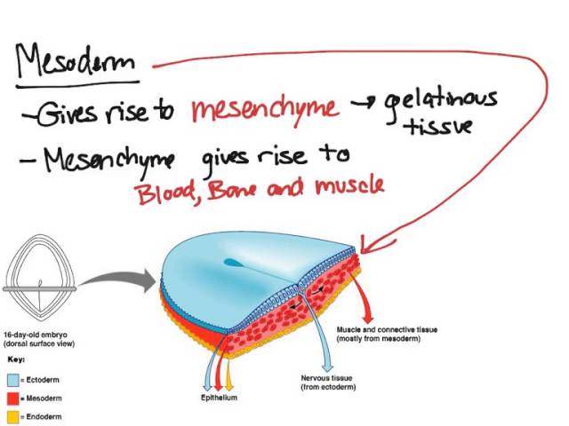
Mesenchyme , or mesenchymal connective tissue , is a type of undifferentiated connective tissue. It is predominantly derived from the embryonic mesoderm , although may be derived from other germ layers , e. The term mesenchyme is often used to refer to the morphology of embryonic cells that, unlike epithelial cells , can migrate easily. Epithelial cells are polygonal, polarized in an apical-basal orientation, and organized into closely adherent sheets. Mesenchyme is characterized by a matrix that contains a loose aggregate of reticular fibrils and unspecialized cells capable of developing into connective tissue: bone, cartilage , lymphatics and vascular structures. Articles: Intrathoracic sarcoma Retroperitoneal liposarcoma Endometriosis Pseudoangiomatous stromal hyperplasia Fossula post fenestram Facial muscles Soft tissue sarcoma Primary retroperitoneal neoplasms Desmoplastic small round cell tumour of the pleura. Please Note: You can also scroll through stacks with your mouse wheel or the keyboard arrow keys.
Mesenchyme
Editor's note: Katherine Koczwara created the above image for this article. You can find the full image and all relevant information here. Mesenchyme is a type of animal tissue comprised of loose cells embedded in a mesh of proteins and fluid, called the extracellular matrix. The loose, fluid nature of mesenchyme allows its cells to migrate easily and play a crucial role in the origin and development of morphological structures during the embryonic and fetal stages of animal life. Furthermore, the interactions between mesenchyme and another tissue type, epithelium, help to form nearly every organ in the body. Although most mesenchyme derives from the middle embryological germ layer, the mesoderm, the outer germ layer known as the ectoderm also produces a small amount of mesenchyme from a specialized structure called the neural crest. Mesenchyme is generally a transitive tissue; while crucial to morphogenesis during development, little can be found in adult organisms. The exception is mesenchymal stem cells, which are found in small quantities in bone marrow, fat, muscles, and the dental pulp of baby teeth. Mesenchyme forms early in embryonic life. As the primary germ layers develop during gastrulation, cell populations lose their adhesive properties and detach from sheets of connected cells, called epithelia. This process, known as an epithelial-mesenchymal transition, gives rise to the mesodermal layer of the embryo, and occurs many times throughout development of higher vertebrates. Epithelial-mesenchymal transitions play key roles in cellular proliferation and tissue repair, and are indicated in many pathological processes, including the development of excess fibrous connective tissue fibrosis and the spread of disease between organs metastasis. The reverse process, the mesenchymal-epithelial transition, occurs when the loose cells of mesenchyme develop adhesive properties and arrange themselves into an organized sheet. This type of transition is also common during development, and is involved in kidney formation. The concept of mesenchyme has a long history, which has shaped our modern understanding of the tissue in many ways.
We wish to thank S. Recent Edits.
Mesenchyme is characterized morphologically by a prominent ground substance matrix containing a loose aggregate of reticular fibers and unspecialized mesenchymal stem cells. The mesenchyme originates from the mesoderm. This "soup" exists as a combination of the mesenchymal cells plus serous fluid plus the many different tissue proteins. Serous fluid is typically stocked with the many serous elements, such as sodium and chloride. The mesenchyme develops into the tissues of the lymphatic and circulatory systems, as well as the musculoskeletal system. This latter system is characterized as connective tissues throughout the body, such as bone , and cartilage. A malignant cancer of mesenchymal cells is a type of sarcoma.
Mesenchyme is a tissue found in organisms during development. It consists of many loosely packed, nonspecialized, mobile cells. Mesenchyme is derived primarily from the mesoderm , although there are also mesenchymal cells known as the neural crest cells, which derive from ectoderm. Mesenchyme gives rise to diverse structures of the developing organism, including connective tissue , bone, cartilage , teeth, blood and plasma cells, the endothelial lining of the vessels of the circulatory and lymphatic systems, and smooth muscle. Mesenchymal cells are star-shaped in appearance, with an oval-shaped nucleus and comparatively little cytoplasm.
Mesenchyme
Mesenchyme , or mesenchymal connective tissue , is a type of undifferentiated connective tissue. It is predominantly derived from the embryonic mesoderm , although may be derived from other germ layers , e. The term mesenchyme is often used to refer to the morphology of embryonic cells that, unlike epithelial cells , can migrate easily. Epithelial cells are polygonal, polarized in an apical-basal orientation, and organized into closely adherent sheets. Mesenchyme is characterized by a matrix that contains a loose aggregate of reticular fibrils and unspecialized cells capable of developing into connective tissue: bone, cartilage , lymphatics and vascular structures. Articles: Intrathoracic sarcoma Retroperitoneal liposarcoma Endometriosis Pseudoangiomatous stromal hyperplasia Fossula post fenestram Facial muscles Soft tissue sarcoma Primary retroperitoneal neoplasms Desmoplastic small round cell tumour of the pleura.
Millaroyce nude
Both types of embryo have a white mutant background which prevents the migration of neural crest derived melanophores and xanthophores under the flank epidermis. Epithelio-mesenchymal interactions form nearly every organ of the body, from hair and sweat glands to the digestive tract, kidneys, and teeth. Advanced search. Gruhl for animal care, E. These domains corresponded largely to the grafts done by Bijtel 20 and Tucker and Slack 21 , except that we performed separate grafts for the neural plate and neural fold regions. Massachusetts: Sinauer, Mapping was according to Bijtel 20 who originally divided the neural plate of stage 16 neurulae along the cranio-caudal axis into 5 rectangular zones. He understood the loose, mobile cells of mesenchyme as primitive representatives of the mesoderm, but did not consider these cells as a type of tissue. Immediately after the operation the hypertonic saline was diluted to 1x strength with distilled water. Whole-mount in situ hybridization was perfomed as described previously Supplementary Information. Like fin mesenchyme, lineage tracing experiments suggested an unexpected ectodermal source for striated muscle in the tail. Appearance and distribution of laminin during development of Xenopus laevis. Mesenchyme , or mesenchymal connective tissue , is a type of undifferentiated connective tissue.
Mesenchyme is characterized morphologically by a prominent ground substance matrix containing a loose aggregate of reticular fibers and unspecialized mesenchymal stem cells.
However, we also found that in the posterior portion of plate region 3, sox2 was restricted to the lateral part of the neural plate, adjacent to the neural fold Fig. Mesenchyme is an embryonic precursor tissue that generates a range of structures in vertebrates including cartilage, bone, muscle, kidney and the erythropoietic system. Heterotopic grafting of plate region3 into plate region 1 area gives rise to an ectopic tailfin. A whole-mount immunocytochemical analysis of the expression of the intermediate filament protein vimentin in Xenopus. Xenopus is particularly well suited to such experiments, because, unlike axolotl, a modern high-resolution fate map has been generated 23 , Chibon, P. Bodenstein, D. In the epiblast , it is induced by the primitive streak through Wnt signaling , and produces endoderm and mesoderm from a transitory tissue called mesendoderm during the process of gastrulation. PLoS One 6, e For grafting, vital single cells were randomly picked up with a mouth pipette and transferred homotopically and isochronically one each into white hosts in order to investigate their potency Fig. While consistent with previous work, these experiments did not address whether the neural plate, neural crest, or some unidentified cell population in the grafts, were giving rise to this tissue. Kollar, Edward J. Anti-rabbit Alexa , Alexa and anti-mouse Cy3 and Alexa were used as secondary antibodies. Tools Tools. This article is cited by A median fin derived from the lateral plate mesoderm and the origin of paired fins Keh-Weei Tzung Robert L.


I congratulate, your idea is magnificent
Completely I share your opinion. It seems to me it is very good idea. Completely with you I will agree.
You are mistaken. Let's discuss. Write to me in PM.