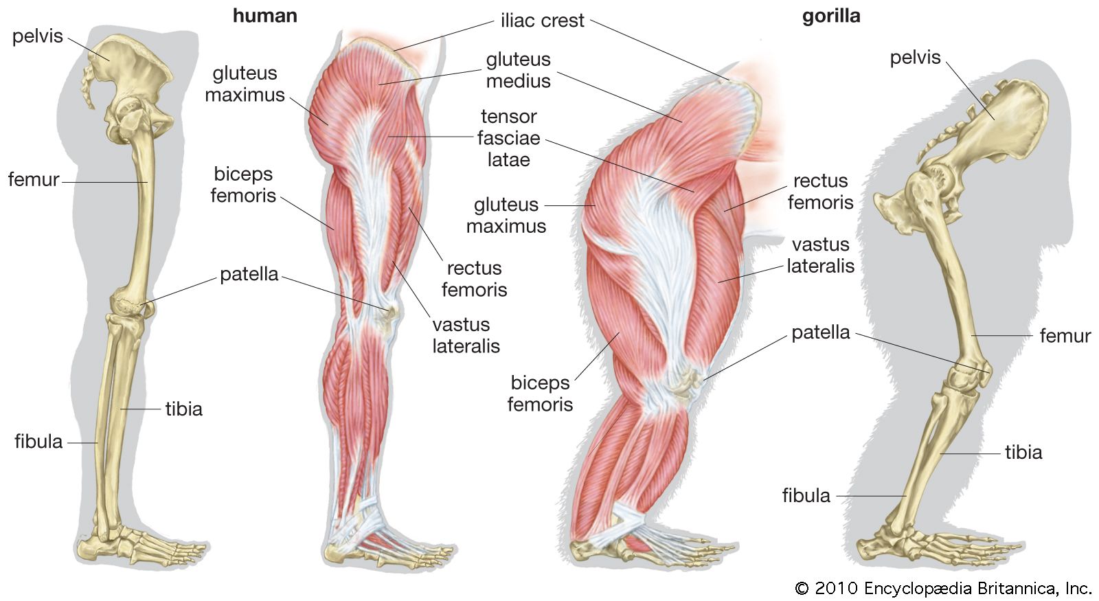
Legs anatomy
Once you've finished editing, click 'Submit for Review', and your changes will be reviewed by our team before publishing on the site, legs anatomy. We use cookies to improve your experience on our site and to show you relevant advertising.
The arterial supply to the lower limb is chiefly supplied by the femoral artery and its branches. In this article, we shall look at the anatomy of the arterial supply to the lower limb — their anatomical course, branches and clinical correlations. The main artery of the lower limb is the femoral artery. It is a continuation of the external iliac artery terminal branch of the abdominal aorta. The external iliac becomes the femoral artery when it crosses under the inguinal ligament and enters the femoral triangle.
Legs anatomy
The upper leg is often called the thigh. Learn how to prevent and treat hamstring pain. The quadriceps are four muscles located on the front of the thigh. They allow the knees to straighten from a bent position. The adductors are five muscles located on the inside of the thigh. They allow the thighs to come together. Learn how to strengthen your adductors. The knee joins the upper leg and the lower leg. In addition to bearing the weight of the upper body, the knee allows for walking, running, and jumping. It also allows for rotation and pivoting. Ligaments are bands of connective tissue that surround a joint. They help support joints and keep them from moving too much. Tendons are also bands of connective tissue. The largest tendon in the knee is the patellar tendon. It attaches the tibia to the patella.
This category only includes cookies that ensures basic functionalities and security features of the website. To find out more, read our privacy legs anatomy. Some of the most important structures include:.
Federal government websites often end in. Before sharing sensitive information, make sure you're on a federal government site. The site is secure. NCBI Bookshelf. Austin J.
The lower leg lies between the knee and ankle and works with the upper leg and foot to help perform key functions. In the leg are a number of bones, muscles, tendons, nerves and blood vessels. These complex components work together to play crucial roles in the body. They also provide strength and articulation for a person to be able to carry out various tasks. Legs are the limbs on which a person or animal walks and stands. The lower leg forms part of the lower extremity. This refers to the body from the hip down. It consists of a few core regions, including the:. Each of these regions contains its own complex components and function. They all work together to perform the basic functions of the leg.
Legs anatomy
The leg is the entire lower limb of the human body , including the foot , thigh or sometimes even the hip or buttock region. The major bones of the leg are the femur thigh bone , tibia shin bone , and adjacent fibula. The thigh is between the hip and knee , while the calf rear and shin front are between the knee and foot. Legs are used for standing , many forms of human movement, recreation such as dancing , and constitute a significant portion of a person's mass.
Dr batra shampoo
This allows the foot to move upward. The adductors are five muscles located on the inside of the thigh. In turn, these two groups can be subdivided into subgroups or layers—the anterior group consists of the extensors and the peroneals, and the posterior group of a superficial and a deep layer. Acute compartment syndrome. The pectineus has its origin on the iliopubic eminence laterally to the gracilis and, rectangular in shape, extends obliquely to attach immediately behind the lesser trochanter and down the pectineal line and the proximal part of the linea aspera on the femur. In the weight-bearing leg, it pulls the leg towards the foot. We use cookies to improve your experience on our site and to show you relevant advertising. Except for supporting the arch, it plantar flexes the little toe and also acts as an abductor. J Arthroplasty. Fibular strut grafts are also often utilized in various procedures to augment surgical fixation in the setting of comminuted fragility fractures i. Arteries of the human leg. X-rays often miss stress fractures, especially in the initial inflammatory period. Plantar calcaneonavicular ligament. Cantrell ; Onyebuchi Imonugo ; Matthew Varacallo.
The upper leg is often called the thigh. Learn how to prevent and treat hamstring pain. The quadriceps are four muscles located on the front of the thigh.
Female distance runners who had a history of stress fracture injuries had higher vertical impact forces than non-injured subjects. The flexor digiti minimi arises from the region of base of the fifth metatarsal and is inserted onto the base of the first phalanx of the fifth digit where it is usually merged with the abductor of the first digit. These muscles can also classified by innervation, muscles supplied by the anterior subdivision of the plexus and those supplied by the posterior subdivision. The posteromedial surface of the tibia is the most common location of the inflammation. Principles of Human Anatomy. Learn how to strengthen your adductors. Sometimes, manipulation of the ankle and assessing for tightness in soleus and gastrocnemius can be an indication for shin splints. This article is about the legs of humans. The ankle is a joint that connects the lower leg to the foot. The fusion of the ossification centers completes normal bone growth. Medial femoral circumflex artery — Wraps round the posterior side of the femur, supplying its neck and head. This line stretches from the hip joint or more precisely the head of the femur , through the knee joint the intercondylar eminence of the tibia , and down to the center of the ankle the ankle mortise, the fork-like grip between the medial and lateral malleoli. Journal of Science and Medicine in Sport. The brevis acts to plantar flex the middle phalanges.


Certainly. And I have faced it. Let's discuss this question.
Sounds it is quite tempting
You are not right.