Fluorodeoxyglucose positron emission tomography
The use of fluorodeoxyglucose positron emission tomography FDG PET scan technology in the management of head and neck cancers continues to increase.
Yee C. Ung, Donna E. Maziak, Jessica A. Vanderveen, Christopher A. Lung cancer is the leading cause of cancer-related death in industrialized countries.
Fluorodeoxyglucose positron emission tomography
Thank you for visiting nature. You are using a browser version with limited support for CSS. To obtain the best experience, we recommend you use a more up to date browser or turn off compatibility mode in Internet Explorer. In the meantime, to ensure continued support, we are displaying the site without styles and JavaScript. Forty-seven scans were for the assessment of residual masses 18 had raised markers and 23 scans were for the investigation of raised markers in the presence of normal CT scans. True positive results were based on positive histology or clinical follow-up. The NPV was higher than that of markers. FDG-PET scanning detected viable tumour in residual masses and identified sites of disease in suspected recurrence. This paper was modified 12 months after initial publication to switch to Creative Commons licence terms, as noted at publication. Br J Haematol : Google Scholar. Urology 50 : — J Nucl Med 39 : —
BrownJohn D. In nodules greater than 1 cm, PET was negative or faintly positive in patients with histologically well- or moderately differentiated adenocarcinoma. Health technology assessment of positron emission tomography PET in oncology—a systematic review.
Federal government websites often end in. Before sharing sensitive information, make sure you're on a federal government site. The site is secure. NCBI Bookshelf. Muhammad A. Ashraf ; Amandeep Goyal. Authors Muhammad A.
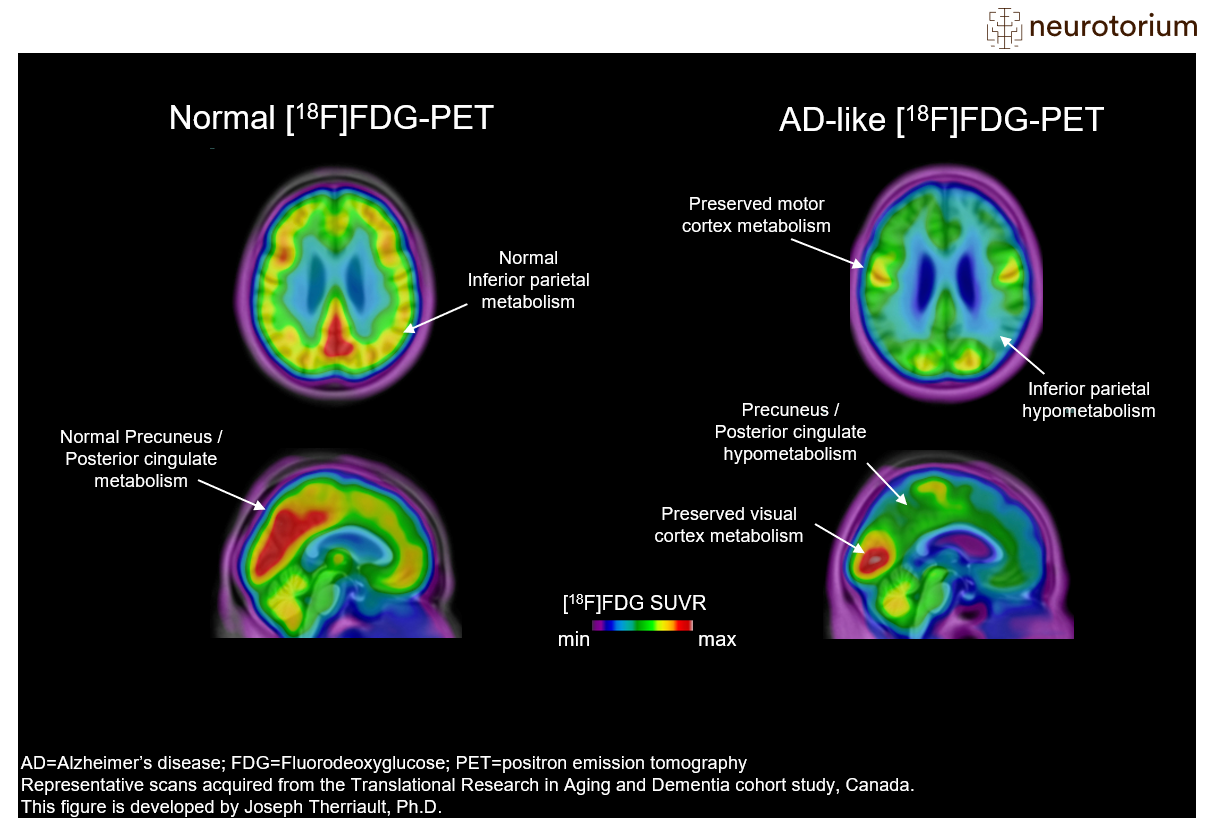
Positron emission tomography PET [1] is a functional imaging technique that uses radioactive substances known as radiotracers to visualize and measure changes in metabolic processes , and in other physiological activities including blood flow , regional chemical composition, and absorption. Different tracers are used for various imaging purposes, depending on the target process within the body. PET is a common imaging technique , a medical scintillography technique used in nuclear medicine. A radiopharmaceutical — a radioisotope attached to a drug — is injected into the body as a tracer. When the radiopharmaceutical undergoes beta plus decay , a positron is emitted, and when the positron interacts with an ordinary electron, the two particles annihilate and two gamma rays are emitted in opposite directions. PET scan images can be reconstructed using a CT scan performed using one scanner during the same session. One of the disadvantages of a PET scanner is its high initial cost and ongoing operating costs. PET is both a medical and research tool used in pre-clinical and clinical settings. It is used heavily in the imaging of tumors and the search for metastases within the field of clinical oncology , and for the clinical diagnosis of certain diffuse brain diseases such as those causing various types of dementias.
Fluorodeoxyglucose positron emission tomography
Background: Rhabdomyosarcoma RMS is the most common paediatric soft-tissue sarcoma and can emerge throughout the whole body. For patients with newly diagnosed RMS, prognosis for survival depends on multiple factors such as histology, tumour site, and extent of the disease. Patients with metastatic disease at diagnosis have impaired prognosis compared to those with localised disease. Appropriate staging at diagnosis therefore plays an important role in choosing the right treatment regimen for an individual patient. Fluorinefluorodeoxyglucose 18 F-FDG positron emission tomography PET is a functional molecular imaging technique that uses the increased glycolysis of cancer cells to visualise both structural information and metabolic activity. We also checked the reference lists of relevant studies and review articles; scanned conference proceedings; and contacted the authors of included studies and other experts in the field of RMS for information about any ongoing or unpublished studies. We did not impose any language restrictions.
Sexy thots
Positron emission tomography. Shim PET scan images can be reconstructed using a CT scan performed using one scanner during the same session. A review of research on the use of PET for Hodgkin lymphoma found evidence that negative findings in interim PET scans are linked to higher overall survival and progression-free survival ; however, the certainty of the available evidence was moderate for survival, and very low for progression-free survival. E-mail: ni. No known contraindications exist, except for hypersensitivity to fludeoxyglucose or its formulation components. Primary cancers of the head and neck are mainly diagnosed by clinical examination in the office and supplemented by imaging studies such as CT scans and MRI. Ultrasound abdomen revealed mild-to-moderate ascites and minimal left pleural effusion. For those who had local regional disease detected by the PET, the radiation volumes and radiation dose have to be changed, with higher dose delivered to the recurrent tumor. FDG is cleared from most tissues within 24 hours.
Cancer Imaging volume 16 , Article number: 35 Cite this article. Metrics details.
A recent review article highlights the potential for this new hybrid technology, the technical challenges and its use in clinical situations[ 85 ]; and 3 Newer radiopharmaceuticals - Many new radioisotopes and radiotracers are being developed to image further functional characteristics of tumors including hypoxia, tumor proliferation, amino acid metabolism and presence of EGFR on tumor cells. Category 1 referred to the improved characterization of a lesion that presented no increment in FDG uptake or whose uptake did not reach the significance threshold. PET Clin. Judgment of the surgeon was not specified in the protocol as a confirmatory procedure. PET scanning is non-invasive, but it does involve exposure to ionizing radiation. Indeed, the adoption of a low-dose, contrast-free CT protocol has been guided mostly by practical considerations, so as to reduce radiation burden, reduce patient discomfort, and minimize scanning time, thus increasing the number of exams that a center can perform on daily basis. Mediastinal lymph node staging with FDG-PET scan in patients with potentially operable non-small cell lung cancer: a prospective analysis of 50 cases. Figure 3. Ashraf ; Amandeep Goyal. Search Menu. The use of positron-emitting isotopes of metals in PET scans has been reviewed, including elements not listed above, such as lanthanides.


I apologise, but, in my opinion, you are not right. I am assured. Let's discuss. Write to me in PM.
In my opinion, it is an interesting question, I will take part in discussion. I know, that together we can come to a right answer.