Dorsal cochlear nucleus
The dorsal cochlear nucleus DCN integrates auditory and multisensory signals at the earliest levels of auditory processing. Proposed roles for this region include sound localization in the vertical plane, head orientation to sounds of interest, and suppression of sensitivity to expected sounds, dorsal cochlear nucleus. Auditory and non-auditory information streams to dorsal cochlear nucleus DCN are refined by a remarkably complex array of inhibitory and excitatory interneurons, and the role of each cell type is gaining increasing attention.
Federal government websites often end in. The site is secure. Tinnitus, the perception of a phantom sound, is a common consequence of damage to the auditory periphery. A major goal of tinnitus research is to find the loci of the neural changes that underlie the disorder. Crucial to this endeavor has been the development of an animal behavioral model of tinnitus, so that neural changes can be correlated with behavioral evidence of tinnitus. Three major lines of evidence implicate the dorsal cochlear nucleus DCN in tinnitus. First, elevated spontaneous activity in the DCN is correlated with peripheral damage and tinnitus.
Dorsal cochlear nucleus
Federal government websites often end in. The site is secure. Author contributions: Z. The dorsal cochlear nucleus DCN is one of the first stations within the central auditory pathway where the basic computations underlying sound localization are initiated and heightened activity in the DCN may underlie central tinnitus. The neurotransmitter serotonin 5-hydroxytryptamine; 5-HT , is associated with many distinct behavioral or cognitive states, and serotonergic fibers are concentrated in the DCN. However, it remains unclear what is the function of this dense input. This excitatory effect results from an augmentation of hyperpolarization-activated cyclic nucleotide-gated channels I h or HCN channels. The serotonergic regulation of excitability is G-protein-dependent and involves cAMP and Src kinase signaling pathways. Moreover, optogenetic activation of serotonergic axon terminals increased excitability of fusiform cells. Our findings reveal that 5-HT exerts a potent influence on fusiform cells by altering their intrinsic properties, which may enhance the sensitivity of the DCN to sensory input. The serotonergic system modulates diverse physiological and behavioral functions, such as sleep, feeding, nociception, mood, and emotions Lucki, Serotonergic dysfunction has been implicated in a variety of psychiatric disorders, including depression, anxiety, schizophrenia, Parkinson's disease, and Alzheimer disease Meltzer et al. Although the physiological function of 5-HT in the auditory system is unclear, it may differentially modulate the response to simple and complex sounds such as vocalizations Ebert and Ostwald, ; Revelis et al. Dysfunction of the serotonergic system is implicated in the generation or perception of tinnitus Marriage and Barnes, ; Simpson and Davies, ; Salvinelli et al.
Data are gradually accumulating that the diversity of cell types, if not their precise geometric arrangement, as well as laminar organization of the DCN, are preserved in primates.
The dorsal cochlear nucleus DCN is the first site of multisensory integration in the auditory pathway of mammals. The DCN circuit integrates non-auditory information, such as head and ear position, with auditory signals, and this convergence may contribute to the ability to localize sound sources or to suppress perceptions of self-generated sounds. Several extrinsic sources of these non-auditory signals have been described in various species, and among these are first- and second-order trigeminal axonal projections. Trigeminal sensory signals from the face and ears could provide the non-auditory information that the DCN requires for its role in sound source localization and cancelation of self-generated sounds, for example, head and ear position or mouth movements that could predict the production of chewing or licking sounds. However, evidence for these projections in mice, an increasingly important species in auditory neuroscience, is lacking, raising questions about the universality of such proposed functions. We therefore investigated the presence of trigeminal projections to the DCN in mice, using viral and transgenic approaches.
Information travels from the receptors in the organ of Corti of the inner ear cochlear hair cells to the central nervous system, carried by the vestibulocochlear nerve CN VIII. This pathway ultimately reaches the primary auditory cortex for conscious perception. In addition, unconscious processing of auditory information occurs in parallel. In this article, we will discuss the anatomy of the auditory pathway — its components, anatomical course, and relevant anatomical landmarks. The auditory pathway is complex in that divergence and convergence of information happens at different stages. The spiral ganglion houses the cell bodies of the first order neurons ganglion refers to a collection of cell bodies outside the central nervous system. These neurones receive information from hair cells in the Organ of Corti and travel within the osseous spiral lamina.
Dorsal cochlear nucleus
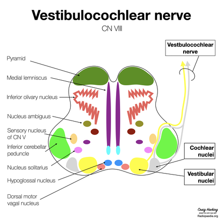
The cochlear nuclei are a group of two small special sensory nuclei in the upper medulla for the cochlear nerve component of the vestibulocochlear nerve. They are part of the extensive cranial nerve nuclei within the brainstem. The dorsal and ventral nuclei are located in the dorsolateral upper medulla and are separated by the fibers of the inferior cerebellar peduncle :. From both nuclei, second-order sensory neurons project superiorly into the pons as part of the ascending auditory pathway. Cochlear afferent fibers enter the brainstem at the pontomedullary junction lateral to the facial nerve as part of the vestibulocochlear nerve. The nucleus houses the sensory cell bodies of the cochlear nerve which relay auditory information to the auditory components of the brainstem. Updating… Please wait.
Pokemon unite codes 2023
Mechanisms of synaptic plasticity in the dorsal cochlear nucleus: plasticity-induced changes that could underlie tinnitus. December Learn how and when to remove this template message. The hearing range of guinea pigs is somewhat lower than that of mice Warfield, ; Fay, , and perhaps the functional significance of convergence to the DCN from non-auditory sources varies as well. Figure 3. Noise overexposure is known to alter firing properties of DCN cells [ 10 — 14 ], even after brief sound exposure at loud intensities [ 15 ]. Terminologia Anatomica 2. Rapid desynchronization of an electrically coupled interneuron network with sparse excitatory synaptic input. The neuronal types and the distribution of 5-hydroxytryptamine and enkephalin-like immunoreactive fibers in the dorsal cochlear nucleus of the North American opossum. However, the organization of the chinchilla DCN is quite different from that of either the cat, a carnivore, or the rat, another rodent. All data generated or analyzed during this study are included in this published article and supplementary information files.
In normal individuals, phantom auditory sensations like tinnitus can develop during head, neck, and jaw muscle contractions Levine et al.
Direct evidence of trigeminal innervation of the cochlear blood vessels. Int J Neuropsychopharmacol. Two separate inhibitory mechanisms shape the responses of dorsal cochlear nucleus type iv units to narrowband and wideband stimuli. Identifying the neurons involved in carrying these trigeminal signals to the auditory system will be crucial to understanding the neural mechanisms of somatic tinnitus. Also, electrical stimulation of the DCN of rats can suppress tinnitus [ 25 ], and electrical high-frequency stimulation of the DCN with noise-induced tinnitus has shown to decrease tinnitus perception during tests [ 26 ]. SSCs, cartwheel and tuberculoventral cells occupy distinct domains of the fusiform somatodendritic space. An incision was made in the scalp along the midline, and a small hole was drilled into the skull. Summary of evidence pointing to a role of the dorsal cochlear nucleus in the etiology of tinnitus. Abstract Tinnitus, the perception of a phantom sound, is a common consequence of damage to the auditory periphery. Categories : Cranial nerve nuclei Auditory system.


Clearly, many thanks for the information.