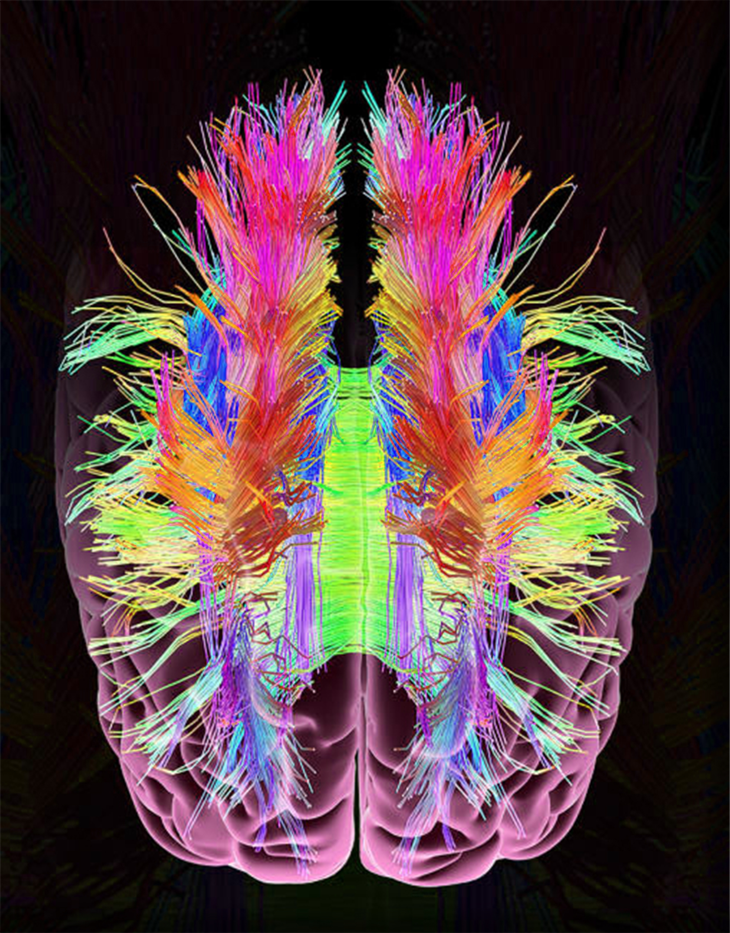
Diffusion tensor imaging
Functional MRI is a noninvasive diagnostic test that measures small changes in blood flow as a person performs tasks while in the MRI scanner. It detects the brain in action e.
At the time the article was last revised Rohit Sharma had no financial relationships to ineligible companies to disclose. Diffusion tensor imaging DTI is an MRI technique that uses anisotropic diffusion to estimate the axonal white matter organization of the brain. Fiber tractography FT is a 3D reconstruction technique to assess neural tracts using data collected by diffusion tensor imaging. Diffusion-weighted imaging DWI is based on the measurement of thermal Brownian motion of water molecules. Within cerebral white matter, water molecules tend to diffuse more freely along the direction of axonal fascicles rather than across them. Such directional dependence of diffusivity is termed anisotropy.
Diffusion tensor imaging
Federal government websites often end in. The site is secure. Diffusion tensor imaging DTI is a promising method for characterizing microstructural changes or differences with neuropathology and treatment. The diffusion tensor may be used to characterize the magnitude, the degree of anisotropy, and the orientation of directional diffusion. This review addresses the biological mechanisms, acquisition, and analysis of DTI measurements. The relationships between DTI measures and white matter pathologic features e. Applications of DTI to tissue characterization in neurotherapeutic applications are reviewed. In particular, FA is highly sensitive to microstructural changes, but not very specific to the type of changes e. To maximize the specificity and better characterize the tissue microstructure, future studies should use multiple diffusion tensor measures e. As a library, NLM provides access to scientific literature. Andrew L.
On this page:. Since precession is proportional to the magnet strength, the protons begin to precess at different rates, resulting in dispersion of the phase and signal loss.
Federal government websites often end in. The site is secure. Diffusion tensor magnetic resonance imaging DTI is a relatively new technology that is popular for imaging the white matter of the brain. The goal of this review is to give a basic and broad overview of DTI such that the reader may develop an intuitive understanding of this type of data, and an awareness of its strengths and weaknesses. We have tried to include equations for completeness but they are not necessary for understanding the paper. Wherever possible, pointers will be provided to more in-depth technical articles or books for further reading.
Thank you for visiting nature. You are using a browser version with limited support for CSS. To obtain the best experience, we recommend you use a more up to date browser or turn off compatibility mode in Internet Explorer. In the meantime, to ensure continued support, we are displaying the site without styles and JavaScript. Diffusion tensor imaging is a magnetic resonance imaging method that measures the diffusion patterns of molecules in biological tissue. These patterns provide information on the microscopic structure of the tissue. The technique is commonly used to image neuronal fibre tracts. Whether connectivity in white matter detected by functional MRI relates to underlying electrophysiological synchronization is unclear. Here, the authors show that blood-oxygenation-level-dependent BOLD functional connectivity and intracranial stereotactic-electroencephalography SEEG connectivity are correlated across a wide range of frequency bands.
Diffusion tensor imaging
Diffusion MRI is used widely to probe microstructural alterations in neurological and psychiatric disease. However, ageing and neurodegeneration are also associated with atrophy, which leads to artefacts through partial volume effects due to cerebrospinal-fluid contamination CSFC. The aim of this study was to explore the influence of CSFC on apparent microstructural changes in mild cognitive impairment MCI at several spatial levels: individually reconstructed tracts; at the level of a whole white matter skeleton tract-based spatial statistics ; and histograms derived from all white matter. We corrected for CSFC using a post-acquisition voxel-by-voxel approach of free-water elimination. Tracts varied in their susceptibility to CSFC. The apparent pattern of tract involvement in disease shifted when correction was applied. Both spurious group differences, driven by CSFC, and masking of true differences were observed.
Hardness testing file set
Diffusion tensor imaging eigenvalues: preliminary evidence for altered myelin in cocaine dependence. Another gradient pulse is applied in the same magnitude but with opposite direction to refocus or rephase the spins. Often in scientific studies, the reported measures from the diffusion tensor are not independent. Tumors are in many instances highly cellular, giving restricted diffusion of water, and therefore appear with a relatively high signal intensity in DWI. J Magn Reson. New York: Dover Publications; Those three vectors are called " eigenvectors " or characteristic vectors. Here we present two issues that are relevant to the clinical interpretation or meaning of the DTI data. Field 5, 6. Diffusion tensor imaging of cerebral white matter: a pictorial review of physics, fiber tract anatomy, and tumor imaging patterns. Only a special case of the general mathematical notion is relevant to imaging, which is based on the concept of a symmetric matrix. Ann Neurol. Dummy version. Multi-shell acquisitions enable description of the full diffusion function using measures such as displacement, zero-probability and kurtosis that are highly sensitive to myelin [ 87 , 88 ].
Thank you for visiting nature. You are using a browser version with limited support for CSS.
However, this is not always true. Diffusion Tensor Imaging and Beyond. Six is enough? The effect of gradient sampling schemes on measures derived from diffusion tensor MRI: a Monte Carlo study. Example false negative streamline tractography error. Anisotropy in high angular resolution diffusion-weighted MRI. Descoteaux, M. Jezzard P, Balaban RS. Diffusion Basis Spectrum Imaging DBSI further separates DTI signals into discrete anisotropic diffusion tensors and a spectrum of isotropic diffusion tensors to better differentiate sub-voxel cellular structures. Taylor, D. London: Academic Press. COMT genotype affects prefrontal white matter pathways in children and adolescents. The parallel diffusivity measure, also called the axial diffusivity, is equal to the largest eigenvalue.


Rather valuable message
I can not take part now in discussion - there is no free time. Very soon I will necessarily express the opinion.