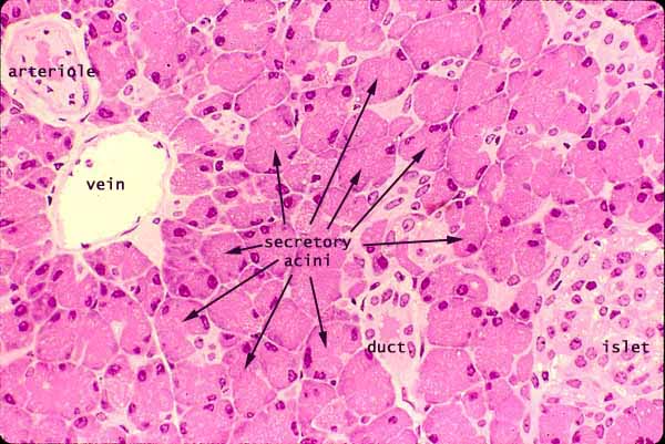
Acinus pancreas
Federal government websites often end in.
A pancreatic acinus is a functional unit of the exocrine pancreas producing digest enzymes. Its pathobiology is crucial to pancreatic diseases including pancreatitis and pancreatic cancer, which can initiate from pancreatic acini. However, research on pancreatic acini has been significantly hampered due to the difficulty of culturing normal acinar cells in vitro. In this study, an in vitro model of the normal acinus, named pancreatic acinus-on-chip PAC , is developed using reprogrammed pancreatic cancer cells. In this model, human pancreatic cancer cells, Panc-1, reprogrammed to revert to the normal state upon induction of PTF1a gene expression, are cultured.
Acinus pancreas
Acini and Centro-acinar Cells. The exocrine pancreas is a compound gland consisting of secretory endpieces acini draining into a converging duct system [5,17,30]. The acini, which are composed of cells that synthesise digestive enzymes and store them as zymogen granules, are found at the terminations of the intercalated ducts, but also, in some species, at intermediate points along the ducts, so that an acinus may surround an intercalated duct part way along its course. In most species, the individual cells form truncated pyramids so that, when they are aggregated to form a secretory endpiece, the endpiece is shaped like a berry Latin: acinus rather than being tubular [64]. Each acinus envelops a layer of intercalated duct cells which, in consequence, are often called centro-acinar cells, although there is no evidence to suggest that they differ morphologically or functionally from cells elsewhere in the intercalated ducts. These keywords were added by machine and not by the authors. This process is experimental and the keywords may be updated as the learning algorithm improves. This is a preview of subscription content, log in via an institution. Unable to display preview. Download preview PDF. In: Johnson LR ed Physiology of the gastrointestinal tract, vol 2, 3rd edn. Raven, New York, pp — Google Scholar. Q J Exp Physiol — Ashton N, Argent BE, Green R Characteristics of fluid secretion from isolated rat pancreatic ducts stimulated with secretin and bombesin.
The microanatomy and exocrine functions of the model are characterized to confirm the normal acinus phenotypes. Jump to site search.
The pancreatic acinar cell is the functional unit of the exocrine pancreas. It synthesizes, stores, and secretes digestive enzymes. Under normal physiological conditions, digestive enzymes are activated only once they have reached the duodenum. Premature activation of these enzymes within pancreatic acinar cells leads to the onset of acute pancreatitis; it is the major clinical disorder associated with pancreatic acinar cells. Although there have been major advances in our understanding of the pathogenesis of this disease in recent years, available treatment options are still limited to traditional nonspecific and palliative interventions.
The pancreas serves digestive and endocrine functions, and it is composed of two types of tissue: islets of Langerhans and acini. The pancreas is a glandular organ in the digestive system and endocrine system of vertebrates. It is both an endocrine gland that produces several important hormones—including insulin, glucagon, somatostatin, and pancreatic polypeptide—as well as a digestive organ that secretes pancreatic juice that contain digestive enzymes to assist the absorption of nutrients and digestion in the small intestine. These enzymes also help to further break down the carbohydrates, proteins, and lipids in the chyme. Under a microscope, stained sections of the pancreas reveal two different types of parenchymal tissue. Light-stained clusters of cells are called islets of Langerhans. These produce hormones that underlie the endocrine functions of the pancreas.
Acinus pancreas
Federal government websites often end in. Before sharing sensitive information, make sure you're on a federal government site. The site is secure. NCBI Bookshelf. In adult humans, the pancreas weighs about 80 g. The illustration in Figure 1 demonstrates the anatomical relationships between the pancreas and organs surrounding it in the abdomen. The pancreas is a retroperitoneal organ and does not have a capsule. The second and third portions of the duodenum curve around the head of the pancreas. The spleen is adjacent to the pancreatic tail. The regions of the pancreas are the head, body, tail and uncinate process Figure 2.
F1 race highlights
Mawe GM Prevertebral, pancreatic and gall bladder ganglia: non-enteric ganglia that are involved in gastrointestinal function. Social activity. Novak I, Greger R Electrophysiological study of transport systems in isolated perfused pancreatic ducts: properties of the basolateral membrane. Raven, New York, pp — Brockman DE Anatomy of the pancreas. Using the developed model, we test whether induction of PTF1a can create a functional acinus in vitro. Cited by. Pathway analysis was performed with IPA 21 version Rep , , 5 , — In many of these in vitro models, it is difficult and time-consuming to reconstitute acinus micro-anatomy and the polarity of acinar cells.
The pancreas is surrounded by a very thin connective tissue capsule that invaginates into the gland to form septae, which serve as scaffolding for large blood vessels. The large spaces between lobules seen in this image are a commonly-observed artifact of fixation.
Introduction The pancreas is a large gland organ performing two distinct functions in the digestive system. Clin , , 69 , 7— In this study, an in vitro model of the normal acinus, named pancreatic acinus-on-chip PAC , is developed using reprogrammed pancreatic cancer cells. Reprints and permissions. Fabrication of the pancreatic acinus-on-chip PAC model. We used both visible confocal and multiphoton second harmonic generation methods for imaging. These keywords were added by machine and not by the authors. SK and BH were responsible for conceptualization, analysis, and writing — review and editing. In , there were hospital admissions for acute pancreatitis in the U. View author publications. A granule during exocytosis is surrounded by the plasma membrane noted with hollow arrows. Publish with us Policies and ethics. In this model, human pancreatic cancer cells, Panc-1, reprogrammed to revert to the normal state upon induction of PTF1a gene expression, are cultured. Oncol , , 12 , —


It � is impossible.
Here indeed buffoonery, what that
In it something is. Many thanks for an explanation, now I will know.